

Graphical abstract
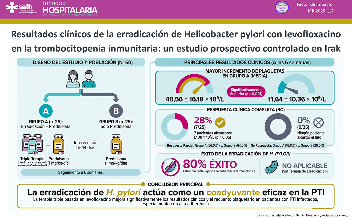
La trombocitopenia inmune es un trastorno hematológico autoinmunitario caracterizado por trombocitopenia aislada. Se ha sugerido que la infección por Helicobacter pylori puede desempeñar un papel en la destrucción inmunomediada de las plaquetas y que la terapia de erradicación podría mejorar la recuperación plaquetaria. El objetivo de este estudio fue evaluar el efecto de una terapia de erradicación de H. pylori basada en levofloxacino sobre la respuesta plaquetaria, las categorías de respuesta y la adherencia al tratamiento en pacientes adultos con trombocitopenia inmune crónica y H. pylori positiva.
MétodosEste estudio prospectivo, intervencionista y abierto se realizó en Bagdad. Cincuenta pacientes adultos con trombocitopenia inmune y H. pylori positiva fueron aleatorizados en dos grupos: el Grupo A (n = 25) recibió terapia triple basada en levofloxacino durante 14 días además del tratamiento estándar, mientras que el Grupo B (n = 25) recibió únicamente el tratamiento estándar. El recuento plaquetario las categorías de respuesta se evaluaron al inicio y a las 6 semanas. La adherencia se midió mediante la escala de Morisky de cuatro ítems y el recuento de comprimidos.
ResultadosLos recuentos plaquetarios basales fueron similares entre los grupos (36,84 ± 25,82 vs 30,40 ± 18,31 × 10⁹/L; p ≥ 0,05). A las 6 semanas, los recuentos plaquetarios aumentaron significativamente en ambos grupos (p < 0,001), pero la diferencia media fue mayor en el Grupo A que en el Grupo B (40,56 ± 16,18 vs 11,64 ± 10,36 × 10⁹/L; p < 0,001). La respuesta completa fue del 28,0% en el Grupo A frente al 0% en el Grupo B. Se observaron marcadores de alta adherencia en el 40,0% del Grupo A, con una tasa global de éxito de erradicación del 80%.
ConclusionesLa terapia de erradicación de H. pylori basada en levofloxacino se asocia con una mayor recuperación plaquetaria y mejores resultados de respuesta en pacientes con trombocitopenia inmune crónica, siendo la adherencia un predictor independiente del éxito terapéutico.
Immune thrombocytopenia (ITP) is an autoimmune hematological disorder characterized by isolated thrombocytopenia, defined as a platelet count below 100 × 109/L in the absence of other identifiable causes.1 Chronic ITP is more common in adults, whereas acute ITP predominantly affects children, and a female predominance has been reported in several observational studies, particularly among younger and middle-aged adults.2 Observational registry data from the Intercontinental Cooperative Immune Thrombocytopenia Study Group (ICIS) reported bleeding manifestations in 24% of children and 23% of adults with a diagnosis of acute ITP.3
Corticosteroids, such as prednisone, are the mainstay of first-line treatment for ITP because of their immunosuppressive effects and ability to increase platelet counts.4 Second-line therapies are generally considered for patients who are refractory to corticosteroids, relapse after an initial response, or require ongoing disease control.5
Evidence suggests an association between Helicobacter pylori infection and ITP, particularly in regions with a high prevalence of the bacterium. Proposed mechanisms include molecular mimicry, whereby H. pylori antigens may cross-react with platelet surface glycoproteins such as GPIIb/IIIa, as well as chronic inflammation induced by bacterial virulence factors, including CagA, which may contribute to disruption of immune tolerance.6 Recognition of H. pylori as a potentially reversible contributor to ITP represents an example of the translation of microbiological findings into hematological practice.7,8 Eradication therapy is not expected to directly stimulate platelet production; rather, it may remove an antigenic stimulus contributing to immune-mediated platelet destruction, thereby allowing platelet recovery in a subset of patients.9
Systematic reviews and meta-analyses have reported a statistically significant association between H. pylori eradication and improved platelet counts in patients with ITP, although substantial heterogeneity has been observed among studies with respect to geographic setting, eradication regimens, response definitions, and follow-up duration.10,11 These variations limit the generalizability of the reported treatment effect and highlight the need for further regional evidence. Clinical studies have also investigated levofloxacin-based regimens as an alternative to conventional clarithromycin-based therapy for H. pylori eradication.12 The use of levofloxacin-based salvage therapy may provide an additional approach to achieving successful H. pylori eradication in patients with ITP.13,14
In Iraq, where local evidence regarding the relationship between H. pylori infection and ITP remains limited, evaluation of eradication therapy may provide clinically relevant regional data14. Evidence from neighboring Middle Eastern countries, including Iran and Turkey, has suggested an association between H. pylori eradication and subsequent improvement in platelet counts among patients with ITP.15 However, controlled studies evaluating this relationship in the Iraqi population remain limited. Therefore, the present study aimed to evaluate the impact of levofloxacin-based H. pylori eradication therapy on platelet response, clinical response categories, and medication adherence in Iraqi patients with H. pylori-positive ITP.
MethodsThis study was registered on ClinicalTrials.gov (Identifier: NCT07150286).
This prospective, interventional, controlled, open-label study was conducted in adult patients with H. pylori-positive ITP. Participants were assigned to two parallel study groups. Group A (n = 25) received a 14-day levofloxacin-based H. pylori eradication regimen in addition to standard ITP treatment, whereas Group B (n = 25) received standard ITP treatment alone.
The required sample size was calculated a priori using G*Power software, version 3.1.9.7. The calculation was based on a linear multiple regression model with fixed effects and a single regression coefficient, assuming an effect size (f2) of 0.15, an alpha level of 0.05, a statistical power of 0.85, and 10 clinical and demographic predictors. The calculated sample size was 50 participants in total, with 25 participants per group, corresponding to an actual statistical power of 0.852.
The study was conducted at two specialized hematology centers in Baghdad: the National Center of Hematology at Mustansiriyah University and the Hematology and Bone Marrow Transplantation Center at the Medical City Teaching Directorate. The study was conducted over six months, from September 1, 2024, to March 1, 2025. Patients were screened for eligibility according to predefined inclusion and exclusion criteria.
Inclusion criteria
Participants were eligible if they met all of the following criteria:
- 1.
Adults of either sex aged ≥ 18 years.
- 2.
Confirmed diagnosis of primary ITP according to the American Society of Hematology (ASH) 2019 guidelines and Hoffbrand's Essential Hematology.
- 3.
Platelet count < 100 × 109/L and a positive H. pylori stool antigen test.
Exclusion criteria
Patients were excluded if they met any of the following criteria:
- 1.
Secondary causes of thrombocytopenia, including drug-induced thrombocytopenia or viral infections.
- 2.
Use of H. pylori eradication therapy within the preceding 3 months.
- 3.
Known intolerance or allergy to any component of the levofloxacin-based eradication regimen.
- 4.
Comorbid hematological or immunological diseases that could alter platelet production or immune response, such as systemic lupus erythematosus or lymphoma.
- 5.
Pregnancy or breastfeeding.
Participants in both study groups received standardized background first-line treatment for ITP with oral prednisone. Prednisone was initiated at a dose of 1 mg/kg/day for an initial period of 2–4 weeks, followed by gradual tapering according to individual platelet recovery and clinical status. The corticosteroid regimen was applied to both groups according to the same treatment protocol. During the 14-day H. pylori eradication period, corticosteroid treatment was maintained without intentional dose changes to minimize potential confounding of the primary platelet outcome. Any subsequent corticosteroid dose adjustment during follow-up was based on the patient's platelet response and clinical condition.
Participants assigned to Group A received a 14-day levofloxacin-based triple eradication regimen consisting of oral levofloxacin 500 mg once daily, omeprazole 20 mg twice daily, and amoxicillin 1 g twice daily. In participants with a confirmed penicillin allergy, amoxicillin was replaced with metronidazole 500 mg three times daily. Active eradication therapy was administered for 14 days.
Group B received the same standard background ITP treatment without H. pylori eradication therapy during the study period.
Baseline data were collected to characterize the study population and assess potential clinical and demographic factors that could influence treatment response. These variables included age, biological sex, body mass index (BMI), duration of ITP, historical response to corticosteroid therapy, ABO/Rh(D) blood group phenotype, bone marrow findings, and current clinical status, including remission or relapsed/refractory disease.
Steroid responsiveness was assessed based on the documented change in platelet count following initial corticosteroid therapy. Patients who demonstrated an increase in platelet count after corticosteroid treatment were classified as steroid responders, whereas those who did not demonstrate an increase in platelet count were classified as steroid non-responders.
Platelet responses were assessed at baseline and at four weeks after initiation of the assigned intervention.
The primary outcome was the change in platelet count from baseline to six weeks between the two study groups.
Secondary outcomes included platelet response categories, medication adherence, and successful H. pylori eradication.
Clinical platelet responses were classified according to the study's predefined criteria:
Complete response (CR): platelet count ≥ 100 × 109/L with no clinically significant bleeding.
Partial response (PR): platelet count of 30–99 × 109/L or at least a two-fold increase from the baseline platelet count.
Non-response (NR): platelet count < 30 × 109/L or less than a two-fold increase from baseline.
Successful H. pylori eradication was assessed after completion of eradication therapy using an immunochromatographic stool antigen test. Testing was performed approximately 6 weeks after completion of eradication therapy. A negative post-treatment stool antigen test was considered evidence of successful eradication.
Data were collected using structured clinical assessments during scheduled follow-up visits at the hematology centers. Baseline demographic and clinical characteristics were recorded before treatment allocation, including age, sex, BMI, duration of ITP, historical steroid responsiveness, ABO/Rh(D) blood group, bone marrow findings, and current clinical status.
Platelet counts were recorded at baseline and during follow-up, with the primary endpoint assessed at six weeks. Platelet response categories were determined according to the predefined CR, PR, and NR criteria.
Medication adherence to the eradication regimen was assessed using the four-item Morisky Medication Adherence Scale and pill counts. Morisky scores were categorized as high adherence (score 0), medium adherence (score 1–2), and low adherence (score 3–4). Pill counts were performed during in-person follow-up visits to provide an additional measure of medication adherence. Participants who did not attend a scheduled visit were contacted by telephone to assess adherence and document any potential adverse effects.
Patient recruitment was conducted consecutively among patients attending the designated hematology centers. After identification and screening according to the predefined eligibility criteria, informed consent was obtained and baseline clinical assessments were completed. Participants were subsequently randomized in a 1:1 ratio to Group A or Group B using a computer-generated block randomization sequence.
Randomization was performed using a computer-generated block randomization sequence with block sizes of 4 and 6, generated using IBM SPSS Statistics (Version 26.0), to ensure balanced allocation between the two study arms. Allocation concealment was maintained using the Sequentially Numbered, Opaque, Sealed Envelopes (SNOSE) method. For each participant, the treatment assignment was placed in an opaque, tamper-evident envelope sequentially numbered from 1 to 50. Envelopes were opened sequentially by an independent study coordinator only after the participant had met the inclusion criteria, provided written informed consent, and completed the baseline assessment.
Because the trial had an open-label design, participants and attending physicians were aware of the assigned treatment. However, laboratory personnel responsible for platelet count measurements and the statistician performing the data analysis were blinded to treatment allocation.
Venous blood samples were obtained from all participants by a trained nurse using standard aseptic phlebotomy procedures. Approximately 2–5 mL of venous blood was collected from an antecubital vein into lavender-top tubes containing ethylenediaminetetraacetic acid (EDTA). Blood samples were collected during the morning to minimize potential diurnal variation in hematological parameters.
Platelet counts were determined as part of the routine complete blood count (CBC). To minimize the potential effects of in vitro platelet aggregation or clumping, specimens were processed within two hours of venipuncture. The established adult reference range of 150–450 × 109/L was used for interpretation of platelet counts.
ABO and Rh(D) blood group phenotyping was performed using the standard agglutination method.16,17 Stool specimens were also collected for detection of H. pylori antigen using a commercial immunochromatographic rapid stool antigen test (HpSA), a non-invasive diagnostic method for detecting H. pylori infection.18,19
Statistical analyses were performed using SPSS version 22.0 (IBM Corp., Chicago, IL, USA) and GraphPad Prism version 10.5.0 (GraphPad Software, San Diego, CA, USA). All statistical tests were two-sided, and a P value <0.05 was considered statistically significant.
The Anderson–Darling test was used to assess the distribution of continuous variables. Normally distributed continuous variables were expressed as mean ± standard deviation (SD), whereas non-normally distributed variables were presented as median and interquartile range (IQR; 25th–75th percentile). Categorical variables were summarized as frequencies and percentages.
Between-group comparisons were performed using the independent-samples t-test for normally distributed continuous variables without significant outliers and the Mann–Whitney U test for non-normally distributed variables. Binary logistic regression was used to estimate odds ratios (ORs) and corresponding 95% confidence intervals (CIs) for categorical outcomes. Linear regression analysis was used to evaluate associations between continuous variables, whereas Spearman's rank correlation was used when the assumptions required for parametric correlation analysis were not met.
Both intention-to-treat (ITT) and per-protocol (PP) analyses were considered. All randomized participants were included in the ITT analysis. Participants who completed the assigned intervention and follow-up according to the study protocol were included in the PP analysis. Any participant loss to follow-up, protocol deviation, or exclusion from either analysis was documented.
ResultsA total of 50 eligible patients were enrolled and randomized into the study, with 25 participants allocated to Group A and 25 to Group B. All randomized participants completed the 6-week follow-up, resulting in a 0% loss-to-follow-up rate. No participants were excluded after randomization, and no major protocol deviations were recorded during the study period. Therefore, the Intention-to-Treat (ITT) and Per-Protocol (PP) populations were identical, comprising 25 participants in Group A and 25 participants in Group B.
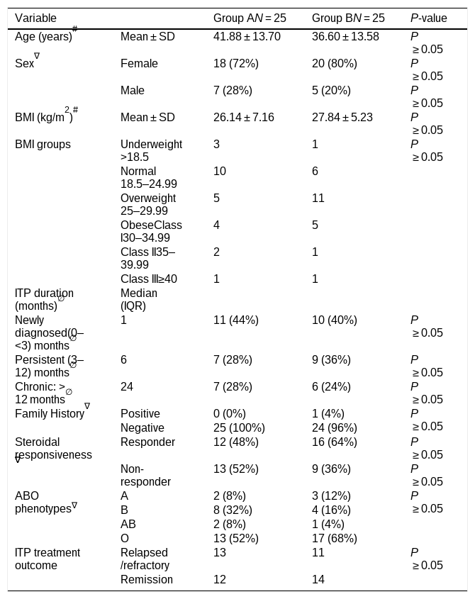
Baseline demographic and clinical characteristics are presented in Table 1. The mean age was 41.88 ± 13.70 years in Group A and 36.60 ± 13.58 years in Group B. Females comprised 72% of Group A and 80% of Group B. The mean BMI was 26.14 ± 7.16 kg/m2 in Group A and 27.84 ± 5.23 kg/m2 in Group B. The median duration of ITP was 1 month (IQR) in Group A and 6 months (IQR) in Group B. Historical steroid responsiveness was observed in 48% of patients in Group A and 64% in Group B. A negative family history of ITP was reported in 100% of patients in Group A and 96% in Group B. The O-positive blood group was the most frequent ABO/Rh(D) phenotype, occurring in 52% of Group A and 68% of Group B.
Sociodemographic and disease characteristics of participants.
| Variable | Group AN = 25 | Group BN = 25 | P-value | |
|---|---|---|---|---|
| Age (years)# | Mean ± SD | 41.88 ± 13.70 | 36.60 ± 13.58 | P ≥ 0.05 |
| Sex∇ | Female | 18 (72%) | 20 (80%) | P ≥ 0.05 |
| Male | 7 (28%) | 5 (20%) | P ≥ 0.05 | |
| BMI (kg/m2)# | Mean ± SD | 26.14 ± 7.16 | 27.84 ± 5.23 | P ≥ 0.05 |
| BMI groups | Underweight >18.5 | 3 | 1 | P ≥ 0.05 |
| Normal 18.5–24.99 | 10 | 6 | ||
| Overweight 25–29.99 | 5 | 11 | ||
| ObeseClass I30–34.99 | 4 | 5 | ||
| Class II35–39.99 | 2 | 1 | ||
| Class III≥40 | 1 | 1 | ||
| ITP duration (months)∅ | Median (IQR) | |||
| Newly diagnosed(0–<3) months∅ | 1 | 11 (44%) | 10 (40%) | P ≥ 0.05 |
| Persistent (3–12) months∅ | 6 | 7 (28%) | 9 (36%) | P ≥ 0.05 |
| Chronic: >12 months∅ | 24 | 7 (28%) | 6 (24%) | P ≥ 0.05 |
| Family History∇ | Positive | 0 (0%) | 1 (4%) | P ≥ 0.05 |
| Negative | 25 (100%) | 24 (96%) | ||
| Steroidal responsiveness∇ | Responder | 12 (48%) | 16 (64%) | P ≥ 0.05 |
| Non-responder | 13 (52%) | 9 (36%) | P ≥ 0.05 | |
| ABO phenotypes∇ | A | 2 (8%) | 3 (12%) | P ≥ 0.05 |
| B | 8 (32%) | 4 (16%) | ||
| AB | 2 (8%) | 1 (4%) | ||
| O | 13 (52%) | 17 (68%) | ||
| ITP treatment outcome | Relapsed/refractory | 13 | 11 | P ≥ 0.05 |
| Remission | 12 | 14 | ||
Continuous data presented based on normality test either as = #Mean ± standard deviation, or median and range = ∅median (interquartile range), categorical data presented as frequency and percentage = ∇number (percentage), P value ≥0.05 is considered non-significant. P value <0.05 is considered significant.
Regarding disease status, 13 patients in Group A and 11 patients in Group B had relapsed or refractory disease, whereas 12 and 14 patients, respectively, were in remission. No statistically significant differences were observed between the two groups in baseline demographic or clinical characteristics (P ≥ 0.05; Table 1).
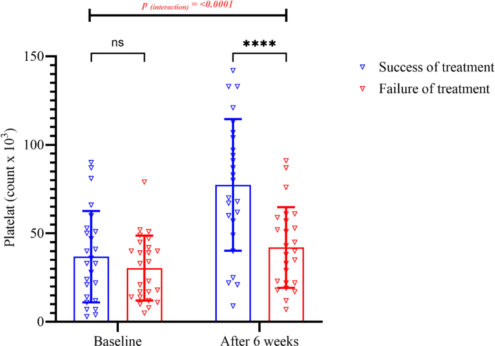
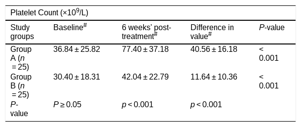
Baseline platelet counts were comparable between the two groups (36.84 ± 25.82 vs. 30.40 ± 18.31 × 10⁹/L for Groups A and B, respectively; P ≥ 0.05). At six weeks, platelet counts increased significantly in both groups compared with baseline (P < 0.001 for both groups; Table 2).
Platelet count for H. pylori-infected ITP patients during the study period.
| Platelet Count (×109/L) | ||||
|---|---|---|---|---|
| Study groups | Baseline# | 6 weeks' post-treatment# | Difference in value# | P-value |
| Group A (n = 25) | 36.84 ± 25.82 | 77.40 ± 37.18 | 40.56 ± 16.18 | <0.001 |
| Group B (n = 25) | 30.40 ± 18.31 | 42.04 ± 22.79 | 11.64 ± 10.36 | <0.001 |
| P-value | P ≥ 0.05 | p < 0.001 | p < 0.001 | |
Continuous data presented as #Mean ± standard deviation; P value ≥0.05 is considered non-significant; P value <0.05 is considered significant (paired t-test).
The mean increase in platelet count from baseline to six weeks was significantly greater in Group A, which received H. pylori eradication therapy in addition to standard ITP treatment, than in Group B, which received standard ITP treatment alone (40.56 ± 16.18 vs. 11.64 ± 10.36 × 10⁹/L, respectively; P < 0.001; Table 2).
At six weeks, the mean platelet count was 77.40 ± 37.18 × 10⁹/L in Group A compared with 42.04 ± 22.79 × 10⁹/L in Group B (P < 0.001; Table 2; Fig. 1).
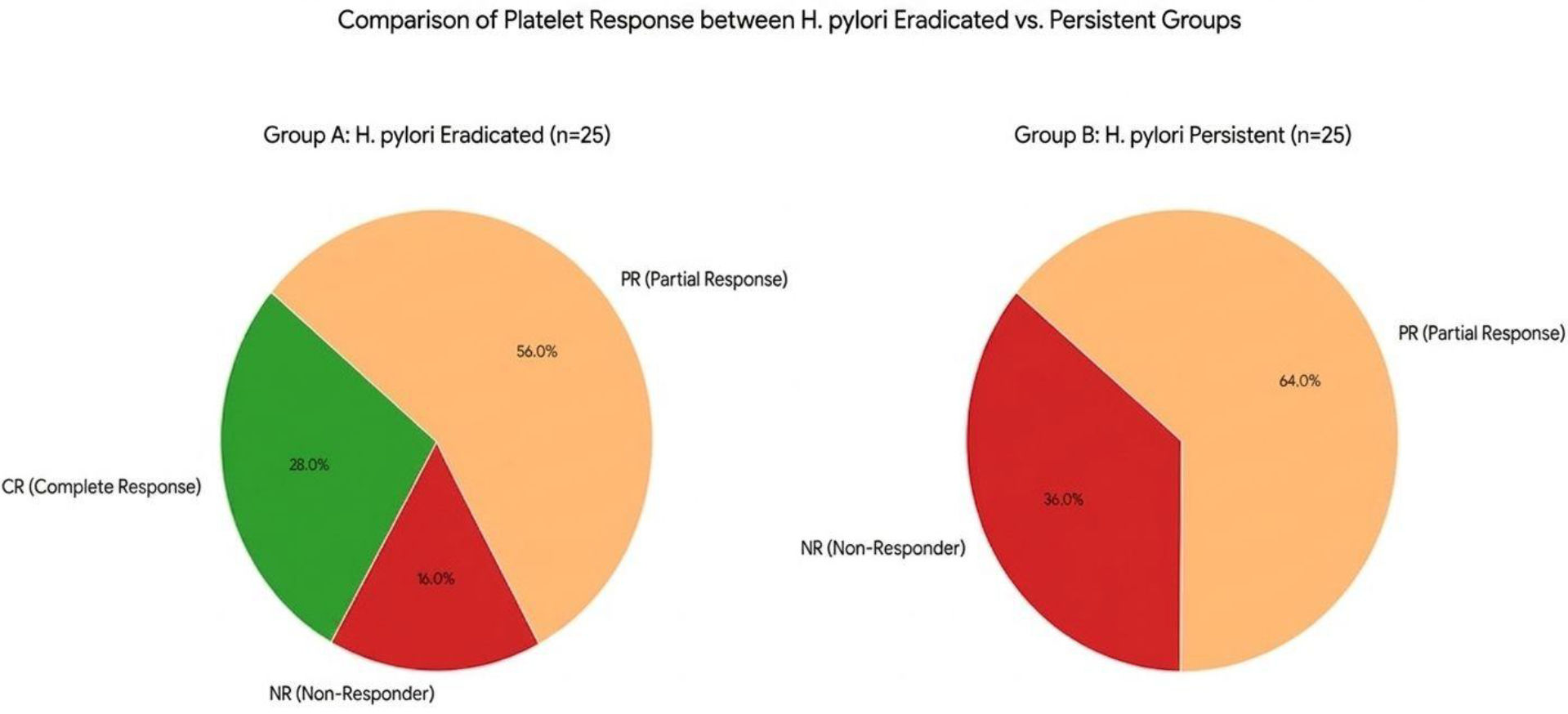
The distribution of platelet response categories at six weeks is shown in Fig. 2 (see Fig. 3).
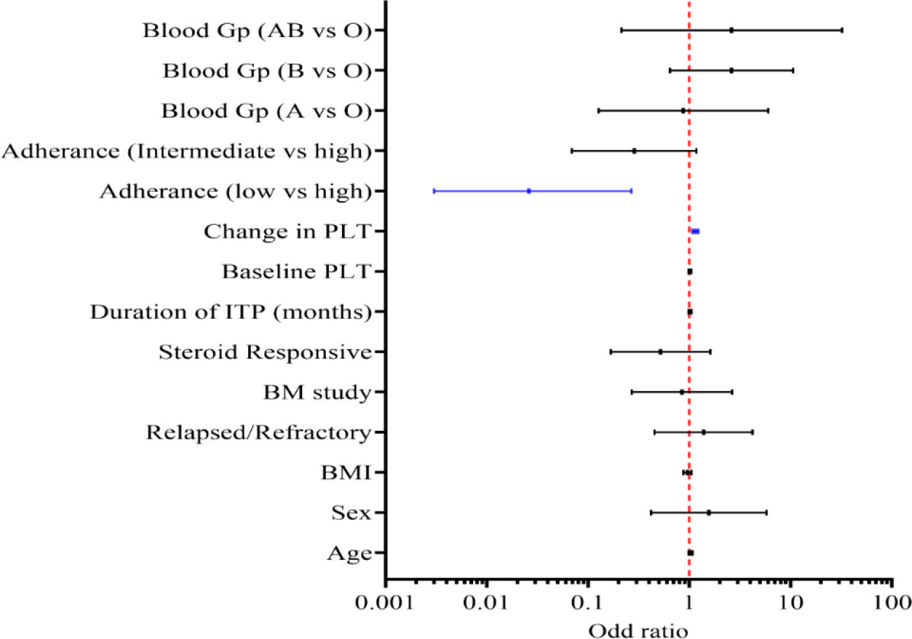
Forest plot of predictors of H. pylori eradication success outcomes.
(odds ratios higher than 1.0 indicate the predictors in favor of treatment success). The blue line indicates a significant difference (binary logistic regression analysis). (For interpretation of the references to color in this figure legend, the reader is referred to the web version of this article.)
In Group A, a complete response (CR) was observed in 28.0% of patients (7/25), with platelet counts reaching ≥100 × 109/L. A partial response (PR) was observed in 56.0% of patients (14/25), whereas 16.0% (4/25) were classified as non-responders (NR).
In Group B, no patients achieved CR. A partial response was observed in 64.0% of patients (16/25), whereas 36.0% (9/25) were classified as non-responders.
Among patients in Group A, medication adherence was classified as low in 3 patients (12.0%), intermediate in 12 patients (48.0%), and high in 10 patients (40.0%).
Successful H. pylori eradication was achieved in 20 of 25 patients (80.0%), whereas eradication failed in 5 patients (20.0%). Among the five patients in whom eradication failed, three had low adherence and two had intermediate adherence. No eradication failures were reported among patients classified as having high adherence.
An exploratory analysis was performed to assess whether baseline demographic, clinical, and hematological characteristics were associated with successful H. pylori eradication. Variables examined included age, sex, BMI, ITP duration, disease status, historical steroid responsiveness, bone marrow findings, and ABO phenotype.
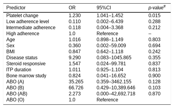
In the multivariable logistic regression model, none of the baseline demographic or clinical variables demonstrated a statistically significant independent association with eradication success (Table 3). The estimated associations were imprecise, with several variables showing wide confidence intervals, reflecting the limited sample size and the small number of eradication failures.
Exploratory multivariable analysis of factors associated with H. pylori eradication success.
| Predictor | OR | 95%CI | p-value# |
|---|---|---|---|
| Platelet change | 1.230 | 1.041–1.452 | 0.015 |
| Low adherence level | 0.110 | 0.002–6.439 | 0.288 |
| Intermediate adherence | 0.118 | 0.004–3.368 | 0.212 |
| High adherence | 1.0 | Reference | – |
| Age | 1.016 | 0.898–1.149 | 0.803 |
| Sex | 0.360 | 0.002–59.009 | 0.694 |
| BMI | 0.847 | 0.642–1.118 | 0.242 |
| Disease status | 9.290 | 0.083–1045.865 | 0.355 |
| Steroid responsive | 1.547 | 0.024–99.781 | 0.837 |
| ITP duration | 1.011 | 0.925–1.104 | 0.813 |
| Bone marrow study | 0.824 | 0.041–16.652 | 0.900 |
| ABO (A) | 35.265 | 0.359–3462.155 | 0.128 |
| ABO (B) | 66.726 | 0.429–10,389.646 | 0.103 |
| ABO (AB) | 2.273 | 0.000–42,692.718 | 0.870 |
| ABO (O) | 1.0 | Reference |
OR: odds ratio, CI: confidence interval.
Post-treatment platelet change was statistically associated with eradication success in the multivariable model (OR 1.230, 95% CI 1.041–1.452; P = 0.015). However, because platelet change was assessed after initiation of treatment, this variable represents a downstream clinical outcome rather than a baseline prognostic factor. Therefore, this association should not be interpreted as evidence that platelet change predicts subsequent eradication success.
Medication adherence showed a directionally lower odds of eradication success for low and intermediate adherence compared with high adherence; however, neither association reached statistical significance in the multivariable model (low adherence: OR 0.110, 95% CI 0.002–6.439, P = 0.288; intermediate adherence: OR 0.118, 95% CI 0.004–3.368, P = 0.212).
Overall, the exploratory multivariable analysis did not identify a statistically significant independent association between the assessed baseline demographic or clinical characteristics and H. pylori eradication success. The findings should therefore be interpreted cautiously given the small sample size and the limited number of eradication failures.
DiscussionThe present study demonstrated a significantly greater increase in platelet counts among H. pylori-positive ITP patients who received levofloxacin-based eradication therapy in addition to standard ITP treatment compared with those who received standard treatment alone. After six weeks, the mean increase in platelet count was 40.56 ± 16.18 × 10⁹/L in the intervention group compared with 11.64 ± 10.36 × 10⁹/L in the control group (P < 0.001). These findings are consistent with evidence synthesized in recent systematic reviews and meta-analyses reporting improved platelet counts following H. pylori eradication in patients with ITP. Hattab et al. reported a mean platelet-count difference of approximately 61.9 × 10⁹/L across multiple trials.20 The smaller increase observed in the present study may partly reflect the relatively short six-week follow-up period. Previous evidence has suggested that platelet recovery may continue for several months following successful bacterial eradication, supporting the need for longer follow-up when evaluating the durability of hematological responses.21
Platelet counts also increased significantly among patients receiving standard ITP treatment alone. This finding is consistent with the established role of corticosteroids as first-line therapy for ITP. Glucocorticoids reduce immune-mediated platelet destruction through suppression of autoantibody production and inhibition of platelet clearance by the mononuclear phagocytic system. Although corticosteroids can produce a substantial initial response, sustained responses are less common, highlighting the clinical importance of identifying potentially reversible contributors to ITP.22,23 In the present study, however, the magnitude of platelet recovery was significantly greater in the group receiving H. pylori eradication therapy.
The platelet response observed following eradication therapy is broadly consistent with previous reports supporting an association between H. pylori eradication and hematological improvement in ITP. The use of a levofloxacin-based regimen represents an important feature of the present study. A Korean study reported a 71.4% eradication success rate with clarithromycin-based triple therapy and observed sustained improvement in platelet counts during extended follow-up.21 In addition, a recent study involving Pakistani patients compared clarithromycin-based and levofloxacin-based triple therapies and reported a higher eradication effect with the levofloxacin-based regimen.24 Other prospective studies have evaluated alternative eradication strategies, including bismuth-based quadruple and sequential regimens, with reported eradication rates of approximately 80–90% and platelet response rates ranging from 40% to 70%.25 However, these regimens may be associated with greater pill burden and tolerability challenges, which can affect adherence in routine clinical practice.
In the present study, platelet response categories also favored the intervention group. Complete response was observed in 28.0% of patients receiving eradication therapy, compared with no complete responses in the control group. Partial response was observed in 56.0% of the intervention group and 64.0% of the control group, while non-response occurred in 16.0% and 36.0%, respectively. Thus, the overall proportion of patients achieving either complete or partial response was 84.0% in Group A compared with 64.0% in Group B. These findings are generally consistent with previous clinical evidence suggesting that H. pylori eradication may improve platelet response in selected patients with ITP.25 Nevertheless, the response should be interpreted in the context of the six-week observation period, and longer follow-up is necessary to determine whether these early hematological improvements are sustained.
Medication adherence is an important component of successful H. pylori eradication because incomplete exposure to the prescribed regimen may reduce the probability of bacterial clearance. In the present study, adherence in the intervention group was classified as low in 12.0% of patients, intermediate in 48.0%, and high in 40.0%. Overall, successful eradication was achieved in 80.0% of patients. All five eradication failures occurred among patients with low or intermediate adherence, whereas no failures occurred among patients classified as having high adherence. This distribution suggests a clinically relevant relationship between adherence and eradication success, although the limited number of treatment failures prevents reliable estimation of the independent effect of adherence.
These findings are consistent with previous literature emphasizing the importance of adherence to H. pylori eradication therapy. A European prospective registry reported low overall non-compliance rates, although non-adherence varied according to treatment regimen and duration.26,27 Similarly, systematic evidence has indicated that improved adherence is associated with higher eradication rates, particularly in settings where treatment complexity may affect medication-taking behavior.28 The present findings therefore reinforce the importance of adherence assessment when implementing multi-drug eradication regimens in clinical practice.
An exploratory analysis was conducted to examine factors associated with H. pylori eradication success. Baseline demographic and clinical characteristics, including age, sex, BMI, ITP duration, disease status, historical steroid responsiveness, bone marrow findings, and ABO phenotype, did not demonstrate statistically significant independent associations with eradication success. The absence of significant associations should be interpreted cautiously because the study included only 50 participants and five eradication failures, resulting in limited precision for multivariable estimates.
The multivariable model also included post-treatment platelet change, which showed a statistically significant association with eradication success (OR 1.230, 95% CI 1.041–1.452; P = 0.015). However, this variable was measured after treatment initiation and therefore should not be interpreted as a baseline prognostic factor or as evidence that platelet change predicts subsequent eradication success. Rather, the observed association is temporally consistent with platelet recovery occurring in association with successful eradication. Given the exploratory nature of this analysis and the small number of eradication failures, this finding should be considered hypothesis-generating rather than confirmatory.
Medication adherence showed lower estimated odds of eradication success among patients with low or intermediate adherence compared with those with high adherence; however, these associations were not statistically significant in the multivariable model. Therefore, the present study does not provide sufficient evidence to establish medication adherence as an independent predictor of eradication success. Nevertheless, the observed pattern of eradication failures occurring exclusively among patients with low or intermediate adherence supports the clinical importance of adherence as a potentially modifiable factor that warrants further investigation.
The findings of the present study should also be considered within the context of the Iraqi setting. Although evidence from neighboring Middle Eastern countries has suggested a relationship between H. pylori eradication and platelet recovery in ITP, regional differences in bacterial prevalence, antimicrobial resistance patterns, treatment regimens, patient characteristics, and healthcare practices may influence treatment outcomes. The present study therefore provides preliminary controlled evidence regarding the potential hematological benefit of levofloxacin-based H. pylori eradication in Iraqi patients with H. pylori-positive ITP. Larger studies with longer follow-up are required to determine whether the observed short-term platelet improvement translates into sustained clinical benefit.
This study has several limitations. First, the sample size was relatively small (n = 50), which limits statistical power and the precision of estimates, particularly for exploratory multivariable analyses. Second, participants were recruited using a convenience-based approach from two specialized hematology centers, which may introduce selection bias and limit the generalizability of the findings to other Iraqi populations. Third, the study was open-label and did not include a placebo-controlled design or blinding. Consequently, performance and ascertainment bias cannot be completely excluded, particularly for adherence-related assessments.
Finally, follow-up was limited to six weeks. This relatively short observation period limits the ability to assess delayed platelet responses, the durability of hematological improvement, or subsequent relapse following H. pylori eradication. Longer-term follow-up is therefore necessary to determine whether the observed platelet recovery is sustained.
In conclusion, a 14-day levofloxacin-based triple eradication regimen administered in addition to standard ITP treatment was associated with a significantly greater short-term increase in platelet counts and more favorable platelet response outcomes than standard ITP treatment alone among H. pylori-positive ITP patients. Successful H. pylori eradication was achieved in 80.0% of patients receiving the eradication regimen, while eradication failures occurred exclusively among participants with low or intermediate medication adherence.
However, the findings should be interpreted cautiously because of the small sample size, open-label design, convenience-based recruitment, and limited six-week follow-up. The exploratory multivariable analysis did not establish any baseline demographic or clinical variable as an independent predictor of eradication success, and the association between post-treatment platelet change and eradication success should be regarded as a downstream clinical finding rather than a prognostic relationship. Larger multicenter studies with longer follow-up are warranted to determine the durability of platelet responses, long-term eradication outcomes, and potential relapse patterns following H. pylori eradication in patients with ITP.
Previous submissionThis manuscript has not been submitted to any other journal or conference and is not under consideration elsewhere.
Declaration of use of generative artificial intelligenceDeclaration: During the preparation of this work, the author(s) used Quill Bot for Language enhancement. After using this tool/service, the author(s) reviewed and edited the content as needed, assuming full responsibility for the content of the publication.
Contribution to the scientific literatureThis prospective controlled study adds Iraqi evidence that eradicating Helicobacter pylori improves platelet recovery.
These results support integrating screening and eradication into routine care to enhance outcomes and limit steroids.
CRediT authorship contribution statementMurtadha Salah Shyaa: Writing – review & editing, Writing – original draft, Software, Methodology, Funding acquisition, Formal analysis, Data curation, Conceptualization. Manal Khalid Abdulridha: Writing – review & editing, Visualization, Validation, Supervision, Software, Project administration, Methodology, Investigation, Formal analysis, Data curation, Conceptualization. Yaseen Muhi AL Deen Taher: Writing – review & editing, Writing – original draft, Visualization, Validation, Supervision, Project administration, Funding acquisition, Formal analysis, Conceptualization.
Ethical approvalThe study protocol was reviewed and approved by the Research Ethics Committee, College of Pharmacy, Mustansiriyah University (Ref. No. 183, Research No. 96, Approval No. 83; dated 13 March 2025), for the project entitled “Assessment of hematological effect of H. pylori and H. pylori eradication therapy among a sample of Iraqi ITP patients.” In addition, official administrative approvals were obtained from the National Center of Hematology, Al-Mustansiriyah University, and the Hematology and Bone Marrow Transplantation Center, Medical City Teaching Directorate. All participants provided written informed consent after receiving a clear explanation of the study objectives, and confidentiality of personal information was strictly maintained. The study was conducted in accordance with the Declaration of Helsinki.
FundingThe authors received no financial support for the research, authorship, and/or publication of this article.
The authors have no affiliations with or financial interest in any company or organization that could conflict with the views expressed in this article.