
To describe the results obtained in terms of effectiveness and safety of intravenous sodium thiosulfate in patients diagnosed with non-uremic calciphylaxis, to identify and analyse possible etiological factors of the disease, and to determine the associated morbidity and mortality.
MethodA multicenter and retrospective study was conducted with patients diagnosed with non-uremic calciphylaxis who received intravenous sodium thiosulfate between 2013 and 2023. Effectiveness was evaluated based on the status of the ulcers at the end of treatment, and safety was assessed according to the main reported adverse effects and the need for dosage adjustment.
ResultsA total of 33 patients from three university hospitals were evaluated (93.9% Caucasian, 78.8% women, mean age 80 [SD 8.1] years) with non-uremic calciphylaxis confirmed by skin biopsy. The localization pattern was 90.9% distal. The following complementary therapeutic measures were undertaken: topical wound care, removal of precipitating factors (mainly vitamin D supplements and vitamin K antagonists), medications to reduce calcification (bisphosphonates, cinacalcet), and techniques to promote ulcer healing. The main associated factors for developing calciphylaxis were: non-uremic chronic kidney disease (81.8%), vitamin D supplementation (72.7%), and hypoalbuminemia (66.7%). The most commonly used sodium thiosulfate dosage was 25 g (n = 26) three times per week (n = 28) intravenously, with a median treatment duration of 11.4 (IQR 5.7-18) weeks. A complete resolution or improvement of the ulcers was achieved in 78.8% of the cases. Adverse effects were observed in 96.7% of patients, with the most common being metabolic acidosis (n = 19) and nausea and/or vomiting (n = 18). Dosage adjustments due to toxicity were necessary in 9% of cases. The significant morbidity rate was 69.7% (n = 23). The main complications were: 57.6% ulcer superinfection and 24.3% poor pain control. The overall mortality rate was 66.7%; 42.4% within the first 6 months after diagnosis and 39.4% secondary to non-uremic calciphylaxis.
ConclusionsSodium thiosulfate shows a potential benefit in the treatment of ulcers due to non-uremic calciphylaxis, with a similar safety profile to that reported for uremic calciphylaxis, considering the high morbidity and mortality associated with the condition. Further studies are needed to determine its efficacy and assess the specific contribution of the different treatments used.
describir los resultados obtenidos en la efectividad y la seguridad del tiosulfato sódico intravenoso en pacientes diagnosticados de calcifilaxis no urémica, identificar y analizar los posibles factores etiológicos de la enfermedad y determinar la morbimortalidad asociada.
Métodoestudio retrospectivo y multicéntrico con pacientes diagnosticados de calcifilaxis no urémica que recibieron tiosulfato sódico intravenoso entre 2013 y 2023. Se evaluó la efectividad según el estado de las úlceras al finalizar el tratamiento y la seguridad según los efectos adversos reportados y la necesidad de ajuste posológico.
Resultadosse evaluaron 33 pacientes de 3 centros hospitalarios (93,9% caucásicos, 78,8% mujeres, edad media 80 [DE 8,1] años) diagnosticados de calcifilaxis no urémica confirmada por biopsia cutánea. El patrón de localización fue 90,9% distal. Se realizaron las siguientes medidas terapéuticas complementarias: curas tópicas, retirada de factores precipitantes (principalmente suplementos con vitamina D y antagonistas de la vitamina K), fármacos para reducir la calcificación (bifosfonatos, cinacalcet) y técnicas para favorecer la curación de las úlceras. Las principales características asociadas a desarrollar la calcifilaxis fueron: enfermedad renal crónica no urémica (81,8%), suplementación con vitamina D (72,7%) e hipoalbuminemia (66,7%). La posología del tiosulfato sódico más utilizada fue 25 g (n = 26) 3 veces por semana (n = 28) por vía intravenosa con una mediana de duración de 11,4 (RIC 5,7-18) semanas. Se obtuvo resolución completa o mejoría de las úlceras en el 78,8% de los casos. El 96,7% de los pacientes presentó efectos adversos, destacando la acidosis metabólica (n = 19) y las náuseas y vómitos (n = 18). El 9% precisó ajuste posológico por toxicidad. La tasa de morbilidad significativa fue del 69,7% (n = 23). Las principales complicaciones fueron: 57,6% sobreinfección de las úlceras y 24.3% mal control del dolor. La tasa de mortalidad global fue del 66,7%; 42,4% durante los primeros 6 meses desde el diagnóstico y 39,4% secundaria a calcifilaxis no urémica.
Conclusionesel tiosulfato sódico muestra un posible beneficio en el tratamiento de las úlceras por calcifilaxis no urémica, con un perfil de seguridad similar al reportado para la calcifilaxis urémica, considerando la elevada morbimortalidad asociada a la enfermedad. Son necesarios estudios adicionales para determinar su eficacia y evaluar la contribución específica de los distintos tratamientos utilizados.
Calciphylaxis is a rare, multifactorial disease characterised by painful skin lesions resulting from calcification and hyperplasia of the middle layer of arterioles and small arteries, leading to ischemia, necrosis, and tissue damage.1,2 It primarily develops in patients with advanced chronic kidney disease (CKD) (stage 4 or higher), including renal transplant recipients and patients on dialysis or pre-dialysis, when it is termed uremic calciphylaxis (UC). However, it has also been documented in patients with normal renal function or in the early stages of CKD, when it is termed nonuremic calciphylaxis (NUC).3–5
The pathogenesis, aetiology, and treatment of NUC are not well established.4 A better prognosis has been observed compared to patients with UC, with an annual mortality rate ranging from 25% to 45%, with sepsis being the leading cause of death.6,7 The optimal therapeutic approach includes pain management, lesion care, elimination of potential precipitating factors, and specific pharmacological treatment.2,3,7 Sodium thiosulfate (STS) is often used off-label to treat calciphylaxis, based on data obtained from retrospective studies of patients with UC.8–12 The mechanism of action of STS is unknown, although several pathways have been proposed to explain its effect, including the formation of soluble complexes with calcium, the restoration of endothelial function, the direct inhibition of calcification, and possible vasodilator and antioxidant effects.13 Adverse effects (AEs) include nausea and vomiting, headache, metabolic acidosis, hypernatremia, volume overload, hypotension, rhinorrhoea, headache, neurological effects (metallic taste, periorbital tingling, decreased hearing), weakness, and hypocalcaemia with a prolonged QT interval.8–10 There are no commercial presentations of intravenous STS in Spain. It is only available in its master formulation. Although there is no standardised intravenous dose of STS for treating calciphylaxis, the most commonly reported dosage guidelines in the literature range from 5 g to 25 g, administered 2–5 times per week.8–11,13
The primary objective of this study was to examine the effectiveness and safety outcomes of 25% STS administered intravenously in patients diagnosed with NUC. Secondary objectives were to identify and analyse potential predisposing factors for NUC and to determine the morbidity and mortality rates associated with the disease.
MethodsDesignA retrospective, multicentre study across 3 university hospitals in Catalonia (Spain). We included patients diagnosed with NUC—as confirmed by skin biopsy—who received intravenous STS between 2013 and 2023 in the internal medicine, nephrology, infectious disease, geriatrics, or dermatology departments, either during hospitalisation or at the daycare hospital. Patients who were lost to follow-up due to transfer to another hospital centre were excluded.
Prior to initiating STS, we collected demographic data, pathological history, calciphylaxis location patterns, and analytical parameters, as well as treatment-related variables, including dosage, frequency, duration, AEs, and concomitant treatments. The calciphylaxis location patterns were classified into 2 groups: distal (lower extremities, fingers and hands) and proximal (trunk, inner thighs, buttocks). Complementary therapeutic measures were documented and categorised into 3 main areas: lesion care, calcification reduction treatment, and elimination of potential precipitating factors. Data collection was based on reviews of the patients' medical histories and electronic prescriptions.
The effectiveness of treatment with STS was assessed based on the clinical evaluation of the treating physician, as documented in the medical record. For data analysis, lesion status after treatment was classified into 4 levels: complete healing (disappearance of the lesion and associated symptoms); improvement (significant reduction in lesion size, formation of granulation tissue, absence of signs of infection or decrease in exudate or pain); stability (no significant change); and deterioration (increase in size, exudate, and pain, or signs of infection). According to clinical assessment, treatment was considered effective when there was complete healing or improvement.
Safety was determined based on the main AEs documented in the clinical history and the need to adjust the treatment dosage.
The identification and analysis of potential predisposing factors were based on the patients' baseline characteristics, analytical parameters, and concomitant treatments described in previous literature as possible risk factors for developing NUC.3,4,6,7,14
Three types of morbidity and mortality rates associated with NUC were determined: significant morbidity, defined as the occurrence of relevant complications during the course of the disease; early mortality, defined as patient death within 6 months of the initial diagnosis of NUC; and mortality related to NUC, characterised by death due to complications associated with disease progression.
Sodium thiosulfate was used off-label in accordance with an institutional protocol that was approved by the pharmacy and therapeutics committee and the medical management of each hospital. In addition, clinical justification was documented in the patient's medical record, and consent was obtained.
This study was authorised as an observational, retrospective study involving medications by the Research Ethics Committee for Medicines of the Fundació Privada Hospital Asil de Granollers. Given the observational nature of the study with no interventions, a waiver of informed consent was approved. The study fully complied with Organic Law 3/2018 of 5 December on Personal Data Protection and the Guarantee of Digital Rights, the fundamental principles of the Declaration of Helsinki, and the Standards of Good Clinical Practice.
Statistical analysisDescriptive statistical analysis was performed for all variables. Continuous variables are expressed by number of patients (n), mean, standard deviation (SD), median, and interquartile range (IQR). Given the inherent limitations due to the small sample size, no formal normality tests were performed to assess the data distribution. Data are expressed by comparing the mean and median. In cases where notable discrepancies were observed, data are described using the median and IQR. Categorical variables are expressed as absolute and relative frequencies for each category. Given the inherent limitations due to the small sample size, no tests were performed for comparative studies. Missing values were recorded for each variable while excluding those with more than 10% missing data. Statistical analysis was performed in accordance with the principles specified in the ICH E9 Guidelines, as well as all Standards of Good Clinical Practice. Statistical analyses were performed using Statistical Analysis System v9.4 (SAS Institute, Cary, USA).
ResultsA total of 33 patients were included. Table 1 shows the demographic data and baseline characteristics of the patients. The lesion location pattern was distal in 90.9% of patients (n = 30) and proximal in 9.1% (n = 3). The diagnosis was confirmed by skin biopsy in all patients.
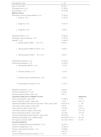
Demographic data and baseline characteristics of patients at the time of diagnosis of nonuremic calciphylaxis.
| Demographic data | n = 33 | |
|---|---|---|
| Age, y, mean (SD) | 80 (8.1) | |
| Caucasian race, n (%) | 31 (93.9) | |
| Sex (femalea), n (%) | 26 (78.8) | |
| Medical history | ||
| Nonuremic chronic kidney disease, n (%) | 27 (81.8) | |
| 10 (37.0) | |
| 10 (37.0) | |
| 7 (26.0) | |
| Diabetes mellitus, n (%) | 18 (54.5) | |
| Peripheral vascular disease, n (%) | 17 (51.5) | |
| Obesityb, n (%) | 14 (42.4) | |
| 6 (46.1) | |
| 4 (30.8) | |
| 3 (23.1) | |
| Hepatobiliary disease, n (%) | 10 (30.3) | |
| Autoimmune disease, n (%) | 10 (30.3) | |
| 7 (70.0) | |
| 1 (10.0) | |
| 1 (10.0) | |
| 1 (10.0) | |
| Malignant neoplasmc, n (%) | 8 (24.2) | |
| Recurrent hypotensiond, n (%) | 6 (18.2) | |
| Previous trauma at lesion site, n (%) | 2 (6.1) | |
| Laboratory data prior to initiation of STS | Reference | |
| Serum urea (mg/dL), mean (SD) | 57 (29.6) | 17–55 |
| Serum creatinine (mg/dL), mean (SD) | 1 (0.4) | 0.6–1.3 |
| Glomerular filtration rate CKD-EPI (mL/min/1.73m2), mean (SD) | 63.6 (21.9) | >90 |
| Total serum protein (g/dL), mean (SD)i | 6 (0.9) | 6–8.3 |
| Serum albumin (g/dL), mean (SD) | 3.2 (0.6) | 3.4–5.4 |
| Total serum calcium (mg/dL), mean (SD) | 9 (0.6) | 8.5–10.2 |
| Serum phosphate (mg/dL), mean (SD)h | 3.2 (0.8) | 2.5–4.5 |
| Calcium-phosphate product (mg2/dL2), mean (SD) | 29.4 (8.0) | <55 |
| Alkaline phosphatase (U/L), median (IQR)i | 85.5 (69.5–121.0) | 44–147 |
| PTH (pg/mL), median (IQR)h | 63.4 (44.7–88.1) | 10–55 |
| Chronic treatments | ||
| Vitamin D supplements and analoguese, n (%) | 24 (72.7) | |
| Vitamin K antagonists, n (%) | 18 (54.5) | |
| Chronic corticosteroid therapyf, n (%) | 16 (48.5) | |
| Irong,h,i, n (%) | 9 (27.3) | |
| Calcium-based phosphorus chelators, n (%) | 1 (3.0) | |
SD, standard deviation; BMI, body mass index; PTH, parathyroid hormone; IQR, interquartile range; STS, sodium thiosulfate; aPTT, activated thromboplastin time; CKD-EPI, Chronic Kidney Disease Epidemiology Collaboration equation.
Obesity was defined as a BMI ≥30. In cases of missing data, patients with a documented history of obesity in their clinical records were considered obese. The BMI data used to define the degrees of obesity are based on 13 patients.
Presence or a previous history of solid or haematological malignant neoplasms at the time of diagnosis of calciphylaxis.
The most commonly used dose of 25% STS administered intravenously was 25 g (n = 26) per session, followed by 12.5 g (n = 5), and 20 g (n = 2). A total of 84.8% (n = 28) of patients received it 3 times per week, 9.1% (n = 3) once per week, and 6.1% (n = 2) twice per week. Median duration was 11.4 (IQR 5.7–18) weeks. Eleven patients received a combination of intravenous therapy and intralesional STS administration. Table 2 shows the complementary therapeutic measures used to promote lesion healing.
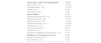
Complementary measures used to promote lesion healing.
| Lesion care | n = 33 | |
| Topical treatmenta, n (%) | 33 (100) | |
| Negative pressure therapy, n (%) | 7 (21.2) | |
| Hyperbaric oxygen therapy, n (%) | 3 (9.1) | |
| Calcification reduction treatment | n = 33 | |
| Bisphosphonate, n (%) | 11 (33.3) | |
| - Alendronate, n (%) | 4 (36.3) | |
| - Risendronate, n (%) | 3 (27.6) | |
| - Pamidronate, n (%) | 2 (18.1) | |
| - Ibandronate, n (%) | 1 (9.0) | |
| - Zoledronic, n (%) | 1 (9.0) | |
| Cinacalcet, n (%) | 4 (12.1) | |
| Elimination of precipitating factors | ||
| Withdrawal of vitamin D supplements and analogues, n (%) | 16 (66.7) | n = 24 |
| Withdrawal of vitamin K antagonists, n (%) | 18 (100.0) | n = 18 |
| Switch to direct-acting anticoagulants, n (%) | 17 (94.4) | |
| Discontinuation of anticoagulant therapy, n (%) | 1 (5.6) | |
| Withdrawal of calcium-based phosphorus chelators, n (%) | 1 (100.0) | n = 1 |
Table 3 shows the effectiveness and safety outcomes of STS treatment. Treatment was considered effective based on the clinical assessment of 78.8% (n = 26) of the patients. However, in 9 of these patients, STS was discontinued before complete healing of the lesions was achieved due to various circumstances: 15.4% (n = 4) died, 11.5% (n = 3) experienced adverse events, and 7.7% (n = 2) experienced events related to venous access. With one exception, all patients experienced AEs related to STS. The reasons for discontinuing STS were as follows: 51.5% (n = 17) lesion healing, 27.3% (n = 9) death, 15.2% (n = 5) AEs, and 6.1% (n = 2) events related to the route of administration (poor venous access and catheter-related bacteraemia leading to endocarditis). Of these patients, 9% (n = 3) required STS dose adjustment due to toxicity; treatment was permanently discontinued in 1 patient, while complete healing was achieved in the other 2 patients.
Effectiveness and safety of sodium thiosulfate therapy.
| Lesion status at the end of STS treatment | n = 33 |
| Improvement, n (%) | 14 (42.4) |
| Complete healing, n (%) | 12 (36.4) |
| Stability, n (%) | 6 (18.2) |
| Deterioration, n (%) | 1 (3.0) |
| Adverse Effects | n = 33 |
| Patients with reported AE, n (%) | 32 (96.7) |
| AE per patient, mean (SD) | 1.4 (0.7) |
| Metabolic acidosis, n (%) | 19 (57.6) |
| Nausea and vomiting, n (%) | 18 (54.5) |
| Hypernatremia, n (%) | 5 (15.2) |
| Volume overload, n (%) | 2 (6.1) |
| Hypotension, n (%) | 2 (6.1) |
| Paroxysmal supraventricular tachycardia, n (%) | 1 (3.0) |
| Modification of dosage due to toxicity | n = 33 |
| Frequency reduction, n (%) | 2 (6.0) |
| Dose reduction, n (%) | 1 (3.0) |
SD, standard deviation; STS, sodium thiosulfate; AE, adverse effects.
The following were identified as the most prominent possible predisposing factors in these patients: nonuremic CKD (81.8%), vitamin D or analogue supplementation (72.7%), hypoalbuminemia (66.7%), hyperparathyroidism (57%), diabetes mellitus (54.5%), use of vitamin K antagonists (54.5%), peripheral vasculopathy (51.5%), chronic corticoid therapy (48.5%), and obesity (42.4%). All patients had more than one concomitant factor.
The rate of significant morbidity was 69.7% (n = 23), with 6 patients experiencing more than 1 complication during the course of the disease. The most relevant complications were as follows: 57.6% (n = 19) lesion superinfection, 24.3% (n = 8) poor pain control, 9% (n = 3) sepsis, 6.1% (n = 2) lesion bleeding, and 3% (n = 1) bacteraemia due to long-term catheter use.
During the study period, total mortality was 66.7% (n = 22). The early mortality rate was 42.4% (n = 14). The NUC-related mortality rate was 39.4% (n = 13). This rate was subclassified into 2 categories: the rate directly attributable to NUC (9.1%, n = 3), caused mainly by sepsis of cutaneous origin; and the rate potentially attributable to NUC (30.3%, n = 10), corresponding to deaths occurring during hospital admission and associated with multiple factors, including deterioration of chronic diseases.
DiscussionThe present study showed that STS in combination with other complementary measures was effective in most patients (78.8%), resulting in complete healing or improvement of lesions, as determined by clinical assessment. However, there are no randomised clinical trials evaluating the efficacy of STS in patients with NUC. Available trials have been conducted exclusively in patients with UC, and their results have not yet been published (NCT03150420, NCT03146793, NCT03319914, NCT05018221). A systematic review by Peng et al. included 358 patients with UC treated with STS. They found an effectiveness rate of 70.1%, defined as an improvement in healing or pain control without associated mortality.15 Nigwekar et al. analysed 172 patients with UC. Of these, 85% completed treatment with STS. Response to treatment was evaluated in 53 patients using a questionnaire sent to treating physicians. A favourable response was observed in 73.6% of patients, broken down as complete healing (26.4%), significant improvement (18.9%) and partial improvement (28.3%).10
However, these results only refer to the use of STS in patients with UC. The available evidence on the use of STS in NUC is limited to isolated cases, case series, or systematic reviews describing its empirical use.4,5,12,16,17 A review by Bajaj et al. included 107 patients with calciphylaxis and normal renal function. No significant association was found between the use of intravenous STS and improvement of skin lesions (hazard ratio [HR] 1.1; 95% confidence interval [CI] 0.6–2.02), or with overall survival (HR 1.01; 95%CI 0.6–1.7).4 However, Altman et al. described a survival rate of 75% in a retrospective analysis of 16 patients with NUC. Patients survived for between 1 and 14 years after diagnosis. The authors attributed this high survival rate to the frequent use of STS in 63% of cases (n = 10).14
In terms of AEs, STS exhibited a safety profile similar to that reported in the literature for both UC and NUC.9 As metabolic acidosis and nausea and vomiting were the predominant AEs, it is important to perform regular laboratory monitoring during treatment and to assess appropriate antiemetic prophylaxis. Reducing the dose or changing the frequency of STS administration could be an option to reduce toxicity. However, in our study, we were unable to determine the optimal strategy for achieving an adequate risk–benefit ratio.
Although current evidence on the treatment of NUC is limited, the use of various strategies has been observed to improve disease progression.2,18 The main complementary measures implemented were topical lesion care and the elimination of precipitating factors. We also employed calcification reduction treatments (bisphosphonates and cinacalcet) and techniques to promote lesion healing (negative pressure therapy and hyperbaric oxygen therapy). In the present study, it was not possible to assess the impact of each of the measures due to the sample size. Bajaj et al. found no association between any combination of medical or surgical interventions (i.e. multimodal treatment) and lesion improvement or survival.4 In contrast, McCarthy et al. found that surgical debridement had a statistically significant benefit in terms of overall mortality.12 Although the magnitude of the effect of the different strategies could not be determined, and despite NUC being a multifactorial condition, the therapeutic management of patients with NUC should be based on applying all available interventions whenever possible.
Nonuremic calciphylaxis was more prevalent among elderly Caucasian women. The predominant characteristics observed in the analysed patient sample correspond to the risk factors for calciphylaxis described in other studies,7 as well as those specifically on NUC.3,4,6,14 However, the conclusions cannot be extrapolated due to the non-population-based design of the study. We draw attention to the normal phosphorus-calcium metabolism, suggesting that mineral abnormalities are not the main aetiological mechanism in NUC. Modifiable etiological factors, such as vitamin D supplements or vitamin K antagonists, should be taken into account. Therefore, when diagnosing patients, it is important to review their pharmacotherapeutic history to evaluate possible alternatives.
The morbidity of NUC was mainly associated with lesion superinfection, poor pain management, and bleeding. Sepsis was the most severe complication. The observed overall NUC-related mortality rate (39.4%) was lower than the rate (52%) reported by Nigwekar et al. in a systematic review of 36 patients with NUC.6 The early mortality rate (41.4%) was also lower than the rate (57.3%) described in the review by McCarthy et al., taking into account that their study also included UC patients.12 As the authors suggested, there appears to be a trend towards longer survival in patients with NUC. They attributed the higher mortality rates among patients with UC to the added risk posed by CKD itself, as well as to the possibility of different risk factors between UC and NUC.12
This study is limited by its retrospective design and heterogeneity in the clinical management of these patients, making it difficult to extrapolate the results. However, to our knowledge, this is one of the longest series published to date in patients with NUC.
In conclusion, the results suggest that STS is of benefit for lesion healing in patients with NUC, and has a similar safety profile to that reported in studies on UC, given the high morbidity and mortality rates associated with the disease. A multimodal treatment approach appears to be a reasonable strategy for NUC. However, further studies are needed to better understand the aetiology of the disease and thus determine the efficacy of the different therapeutic measures used.
Contribution to the scientific literatureNonuremic calciphylaxis is a rare disease, many aspects of which remain unknown, and for which there is little scientific evidence. This study provides real-life data on the effectiveness and safety of using STS off-label to treat NUC.
The results obtained in this multicentre study demonstrate the usefulness of intravenous STS for patients with NUC. They provide relevant information on managing this disease and contribute to optimising the clinical approach.
CRediT authorship contribution statementLaura Cardona-Roca: Writing – review & editing, Writing – original draft, Project administration, Methodology, Formal analysis, Data curation, Conceptualization. Laura Borràs Trias: Writing – review & editing, Validation, Supervision, Methodology, Formal analysis, Conceptualization. Rosario Bueno Uceda: Writing – review & editing, Validation, Data curation. Adrià Siles: Writing – review & editing, Validation, Data curation. Adrián Vilariño Seijas: Writing – review & editing, Validation, Data curation. Nuria Rudi Sola: Writing – review & editing, Validation, Supervision, Methodology, Data curation, Conceptualization. Carlos Seguí-Solanes: Writing – review & editing, Writing – original draft, Validation, Supervision, Methodology, Formal analysis, Conceptualization.
FundingNone declared.
None declared.