

To assess pharmacobezoar formation in a case of intoxication following the massive ingestion of extended-release divalproex sodium tablets, using an in vitro model conducted in the pharmacy laboratory.
MethodsAn in vitro model was created to simulate gastric conditions and evaluate pharmacobezoar formation. Simulated gastric fluid was prepared according to the European Pharmacopeia procedure. The conglomerate's size and pH were measured at 30 min, 1 h, 2 h, 4 h, 8 h, 12 h, and 24 h intervals. Serum valproic acid concentrations were determined at all mentioned time points and then compared with the patient's serum concentrations.
ResultsThe in vitro model revealed the formation of a solid pharmacobezoar (5 cm x 6 cm) after 2 h (pH = 1.5), which remained stable for 24 h. The peak serum concentration of valproic acid occurred approximately 22 h post-ingestion, reaching 656 μg/mL. The in vitro assay indicated a similar peak concentration around 24 h after the tablets were immersed in the simulated gastric fluid.
ConclusionsThe experiment conducted supports the hypothesis of pharmacobezoar formation following excessive ingestion of extended-release divalproex sodium tablets. Understanding which drug formulations can potentially cause pharmacobezoars formation is crucial for toxicological management.
Evaluar la formación de farmacobezoar a propósito de un caso de intoxicación tras la ingestión masiva de comprimidos de divalproato sódico de liberación prolongada, utilizando un modelo in vitro realizado en el laboratorio de farmacotecnia.
MétodosSe desarrolló un modelo in vitro para simular las condiciones gástricas y evaluar la formación de farmacobezoares. El fluido gástrico simulado se preparó siguiendo el procedimiento de la Farmacopea Europea. El tamaño del conglomerado y el pH se midieron a intervalos de 30 min, 1 h, 2 h, 4 h, 8 h, 12 h y 24 h. También se determinaron las concentraciones séricas de ácido valproico en estos mismos intervalos, comparándolas después con los niveles obtenidos en la paciente.
ResultadosEl modelo in vitro reveló la formación de un farmacobezoar sólido (5 cm x 6 cm) después de 2 horas (pH = 1.5), que se mantuvo estable durante 24 horas. La concentración máxima de ácido valproico en suero se alcanzó aproximadamente 22 horas después de la ingestión, alcanzando un valor de 656 μg/mL. El ensayo in vitro indicó una concentración máxima similar alrededor de 24 horas después de que los comprimidos fueran sumergidos en el fluido gástrico simulado.
ConclusionesEl experimento realizado ha permitido respaldar la hipótesis de la formación de farmacobezoar tras la ingestión masiva de comprimidos de divalproato sódico de liberación prolongada. Conocer qué tipo de formulaciones pueden estar implicadas en la formación de farmacobezoares es crucial para su manejo toxicológico.
A frequent cause for hospital emergency admissions is intentional drug overdose.1 In some cases, this can lead to the formation of pharmacobezoars, which are accumulations of undigested or partially digested drugs in the gastrointestinal system and can cause obstructions and serious complications.2,3 Although the exact process of pharmacobezoar formation is not completely understood, it is hypothesised to be the result of a combination of several factors, including medication dosage, the physical and chemical properties of the drug, delays in gastric emptying, and anatomical irregularities in the gastrointestinal tract.3,4 Extended-release (ER) formulations have frequently been related to pharmacobezoar formation. This type of drug is formulated for gradual release of the active ingredient in the gastrointestinal tract over time; some excipients, such as cellulose derivatives, are frequently present in ER formulations and could contribute to tablet adhesion, potentially leading to the formation of a pharmacobezoar, in the case of massive ingestion.5,6 There are several case reports in the literature about pharmacobezoar formation associated with the ingestion of enteric-coated aspirin, sustained-release quetiapine, verapamil, venlafaxine and quinidine, as well as potassium and iron supplements.7
In cases of severe poisoning, gastrointestinal decontamination is a widely used method to reduce absorption of the ingested substance.8 However, the use of activated charcoal and gastric lavage tubes could be clinically ineffective in cases of pharmacobezoars, whose treatment often involves endoscopy to remove the bezoar.3,8 Furthermore, the bezoar results in an extended absorption period, resulting in a delayed attainment of peak plasma concentration. This is, usually accompanied by a prolonged duration of high drug concentrations near the peak, as noted in previous case reports. This can result in delayed toxicity and longer-lasting symptoms.9
Divalproex sodium, a mixture of valproic acid (VPA) and sodium valproate, is a commonly used antiepileptic drug to treat various types of epilepsy, bipolar disorder, and migraines.10 In severe cases, divalproex sodium toxicity commonly manifests as central nervous system depression, while hyperammonemic encephalopathy and hepatotoxicity can pose life-threatening risks.11,12
This study was prompted by a real-life concerning a 16-year-old woman, with a history of mental health issues, who was being treeated with olanzapine, zolpidem, clonazepam, and divalproex sodium ER tablets (Depakine Chrono® 500 mg). She ingested 47 tablets of 500 mg each (23.5 g, 427.27 mg/kg). Upon admission to the emergency department, two hours after the overdose, she underwent gastrointestinal decontamination with activated charcoal. Her initial valproate serum concentration was 154 μg/mL, and a second dose of charcoal was administered 13 hours later when the patient showed worsening symptoms, including decreased consciousness, mydriasis, nausea, vomiting, and choreiform movements. Her valproate serum concentration subsequently increased to 309 μg/mL, and ammonia levels were also elevated. The maximum valproate serum concentration peaked at 656 μg/mL, 22 h post-ingestion, which is later than the usual 10–12 h for delayed-action preparations. This unusual timing coupled with the mild initial symptoms suggested the formation of a pharmacobezoar. This atypical pharmacokinetic profile, along with the progressive toxicity, raised the suspicion of pharmacobezoar formation.
The Pharmacy Compounding Unit conducted an in vitro study at the suggestion of the Toxicology department to investigate whether ER divalproex sodium tablets could aggregate into a solid mass (pharmacobezoar) under gastric conditions. This could potentially explain the delayed absorption and toxicity observed in this case.
MethodsThis model aimed to simulate gastric conditions and study how the tablets might aggregate into solid conglomerates, increasing the risk of delayed toxic effects.
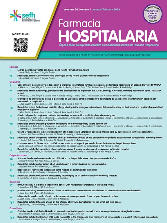
Simulated gastric fluid (SGF) was prepared in the laboratory of the pharmacy department following the European Pharmacopeia procedure, by dissolving 2 g of sodium chloride (Acofarma. Lot. no: 191426-P-1) and 80 mL 1.0 M hydrochloric acid (ITW Reagents. Lot. no: 0002055967) in 1 L of purified water (Fresenius Kabi. Lot. no: 13SIP101).13 The SGF was then poured into a glass container and magnetically stirred at 30 rpm and at a temperature of 37 °C for 24 h. The resulting SGF appeared as a clear and transparent solution with a pH of 1–2 throughout the in vitro assay. This was monitored using pH-indicator strips (Fig. 1(A)).

A total of 47 ER of divalproex sodium (Depakine crono®, Sanofi Aventis S.A. Lot. no: 2R38L) 500 mg tablets (23.5 g), equivalent to the reported ingested dose, were placed inside a 70 cm x 50 cm polyester mesh bag (Fig. 1(B)). The mesh bag was used to simulate the gastric environment by maintaining the tablets in close and continuous contact with the SGF, mimicking their position in the stomach after ingestion. The mesh bag was securely attached to a crystal stick positioned on the top of the glass beaker, ensuring complete immersion of the tablets in the SGF. The formation of the conglomerate was observed by measuring its size at different time intervals (30 min, 1, 2, 4, 8, 12 and 24 h).
The VPA concentration in the solution was measured at the same time intervals in 3 mL aliquots using a turbidimetric immunoassay (Atellica® Solution CH 930). Prior to sampling, the entire SGF solution was agitated for 10 seconds at 150–300 rpm, in order to ensure proper homogenization of the medium and an even distribution of dissolved VPA.
ResultsAfter 2-h immersion in the SGF, the formation of a solid 5 cm x 6 cm pharmacobezoar was observed (pH = 1.5) (Fig. 2(A)). Additionally, Fig. 2(B) shows that a standard 30F orogastric lavage tube could not be used to remove the pharmacobezoar. Over time, the tablets became more adherent to each other, forming a more compact conglomerate. After 24 hours of the experimental procedure, the divalproex sodium tablets were intact (Fig. 3(A)) and had formed a stable pharmacobezoar that was collected in the mesh bag. This remained relatively constant throughout the duration of the assay, and was relatively firm and resistant to mechanical manipulation (Fig. 3(B)).


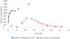
The VPA concentrations obtained in the in vitro model were compared with those reported in vivo (see Fig. 4). VPA was monitored using a validated immunoassay method (Atellica™ CH Valproic Acid, Siemens). As can be seen in Fig. 4, in this patient, the initial blood drug concentration was 154 μg/mL, and after 6 h, it decreased to 81 μg/mL. Twelve hours after ingestion, the serum drug concentration had risen to 309 μg/mL without any further administration, suggesting continuous absorption of divalproex sodium. The peak VPA concentration in serum occurred approximately 22 h after ingestion, measuring 656 μg/mL. The in vitro assay also demonstrated a peak concentration approximately 24 h after the tablets were immersed in the simulated gastric fluid. Guidelines for therapeutic drug monitoring of antiepileptic drugs indicate that the elimination half-life of VPA ranges from 11–12 h, with an increase observed in cases of overdose.12 The fact that the peak concentration occurred at 22 h after ingestion indicates a delay in absorption (“flip-flop absorption”), which could be caused by the formation of a pharmacobezoar.
Discussion
This experiment aims to provide insights into the specific characteristics that could contribute to the formation of pharmacobezoars in cases of divalproex sodium overdose.
An overdose of divalproex sodium can lead to various symptoms, including reduced level of consciousness, hypotension, hypernatremia, and hyperammonaemia, which can result in severe encephalopathy. In severe cases, hyperlacticaemia and metabolic acidosis may occur.14
In this patient, there is a notable discrepancy between the mild initial symptoms experienced and the large quantity of divalproex sodium ingested (>400 mg/kg). The serum VPA concentration measured 6 h after ingestion was 81 μg/mL, which falls within the therapeutic range of 50–100 μg/mL.13 Delayed peak VPA concentrations in the toxic range might not always align with clinical symptoms of toxicity. Ingels et al. conducted a retrospective case series involved 173 intoxicated patients, 15% of these presented concentrations of VPA that were not in the toxic range after an acute overdose.15 Therefore, the initial serum VPA concentrations could be misleading, and suggest several possibilities, such as a smaller ingested dose than the one reported or a potential pharmacobezoar formation. The latter seems the more plausible option in this case, after the in vitro assay was performed.
Delayed peak serum drug concentrations and pharmacobezoar formation have been observed with various ER medications such as paracetamol,16,17 nifedipine,18 venlafaxine,19 quetiapine20 and clomipramine.5,21 ER formulations are designed to maintain stable drug serum concentrations, minimizing adverse effects from high drug concentration and preventing symptom breakthroughs below the therapeutic range.6 To achieve this, ER preparations are designed to retain their size and shape in the gastrointestinal tract for an extended period.3 The ER coating of divalproex sodium tablets typically contains hydroxypropylmethylcellulose, macrogol 6000, talc, titanium dioxide (E-171) and polyacrylate. Under moist conditions, hypromellose or hydroxypropylmethylcellulose can generate a sticky consistency, causing the tablets to adhere to each other and form a relatively solid conglomerate.10 Pharmacobezoar formation can potentially slow down drug release compared to individual units, as the reduced surface-to-volume ratio prolongs dissolution time and delays maximum drug absorption.3 Furthermore, apart from the delayed rise in serum drug concentrations, the risk of complications could be heightened by the gastric or intestinal retention of the pharmacobezoar.3
Standard orogastric lavage tubes have a maximum width of 30-32F, a diameter of approximately 9 mm, and side holes measuring 4–11 mm (30F). Neither the formed pharmacobezoars nor the pharmaceutical preparations in the form of single tablets of divalproex sodium could pass through the openings of a standard 30F orogastric lavage tube.3 Given that the majority of pharmacobezoars consist of sticky gelatinous masses, the impact on passage through the tube is not solely determined by pill size. The pharmacobezoar formed in the in vitro assay measured 5 cm x 6 cm, supporting the fact that in cases of ER formulations that do not disintegrate in the stomach or tend to form bezoars, lavage tubes may be insufficient to remove the entire ingested drug.5
Instances of compelled removal of pharmacobezoars have been recorded through various procedures, including the use of fizzy beverages for ingestion and/or intrabezoar injection,22,23 extraction by endoscopy, whole bowel irrigation,9 sometimes with prior fragmentation5,19,21,24–26 rectal extraction,27 or surgical intervention.19
To address ethical concerns, conducting human research on pharmacobezoars poses challenges, prompting us to opt for an in vitro experimental study. However, it is crucial to acknowledge that the experimental conditions in vitro differ from the dynamic environment of in vivo circumstances. The latter involve continuous secretion of gastric juice, concomitant or subsequent intake of fluids and/or food, pH changes, hormonal release, absorption, digestion, and gastric emptying.
While it may be difficult to replicate these conditions fully in vitro, it is crucial to develop procedures that simulate them to validate and confirm our experiment's findings. Additionally, our experimentation was limited to 24 h, and longer observation periods could have resulted in greater dissolution of the formed clumps. An evaluation of the most effective technique for eradicating pharmacobezoars was not carried out in this in vitro investigation, and it remains unclear whether eliminating established pharmacobezoars improves patient outcomes.
ConclusionIn conclusion, this collaborative experiment between the Pharmacy and Toxicology departments has validated the hypothesis of pharmacobezoar formation following excessive ingestion of ER divalproex sodium tablets. Understanding which drug formulations can potentially cause pharmacobezoars formation is crucial for toxicological management. This enables the appropriate methods for digestive decontamination to be assessed and helps to clarify the time course of drug serum concentrations and the onsetof adverse effects.
Contribution to the scientific literatureThis study contributes scientifically to explore the effects of massive ingestion of extended-release divalproex sodium tablets and the formation of pharmacobezoars. It provides insight into how these tablets can aggregate into solid conglomerates in the stomach, delaying drug release and increasing the risk of delayed toxic effects. The research also enhances the understanding of divalproex sodium intoxication and its abnormal plasma concentration behavior in overdose cases, which is crucial for managing poisoning.
Overall, the study advances knowledge on the pharmacokinetics of extended-release drugs in overdose scenarios and supports the development of simulation models for future research in toxicology and clinical pharmacy.
CRediT authorship contribution statementThais Lizondo López: Writing – review & editing, Writing – original draft, Validation, Methodology, Conceptualization. Silvia González Suárez: Validation, Methodology, Investigation, Conceptualization. Carmen López-Cabezas: Writing – review & editing, Supervision, Methodology, Conceptualization. Judit Julian Peña: Validation, Methodology. NCaira Rico Santana: Validation. Dolors Soy Muner: Supervision, Conceptualization. Núria Fernández Hernández: Validation, Data curation. Emilio Salgado García: Visualization, Validation, Project administration, Methodology, Investigation, Formal analysis, Conceptualization.
Ethical approvalRegarding ethical considerations, it is noteworthy that all methods were carried out in accordance with relevant guidelines and regulations. No biological samples were used in the in vitro study. As for the clinical case, informed consent was obtained from the patient prior to drafting this article. All analytical data reported, including serum valproic acid concentrations, were analyzed as part of the patient's clinical-toxicological follow-up process, following routine practice.
FundingThe authors declare that they received no funding for this study.
The authors declare no conflicts of interest related to this study.